Floaters and Flashes: When Are They an Emergency?
What floaters and flashes are, why they increase with age, who gets them more, when they are an emergency, retinal tears and detachment, migraine flashes, what to do, how they are checked and treated, follow-up and prevention.
By Michel Geha, O.O.D.
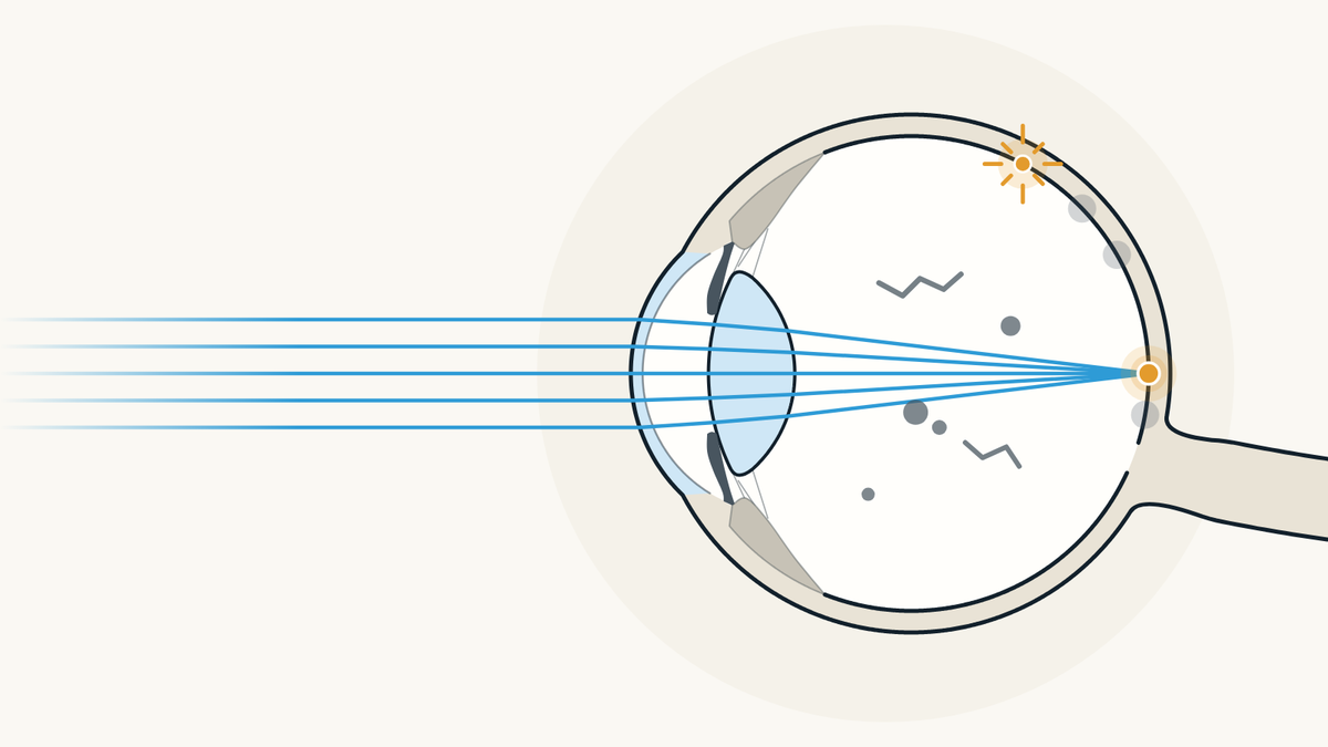
Floaters are small specks or threads that drift across your vision, and flashes are streaks of light you see with no light outside. A few that come with age are usually harmless. It is urgent when many appear at once, or come with flashes, a shadow or a grey curtain over your vision.
What are eye floaters?
They are tiny clumps of gel or cells in the vitreous, the clear gel that fills the eye, and what you see is their shadow on the retina. The American Academy of Ophthalmology (AAO) describes them as specks, dots, circles, lines or cobwebs that seem to be in front of the eye but float inside it.
The Canadian Association of Optometrists (CAO) says they move when your eyes move, with a lag, and seem to dart away when you try to look straight at them. You notice them most against a plain bright background, like a white wall or a blue sky. Almost everyone sees a few at one time or another, the CAO adds.

What causes flashes of light in the eye?
Most often, the vitreous gel rubbing or pulling on the retina. The AAO compares these flashes to lightning streaks or the "stars" you see after a blow to the head. According to the AAO, this light starts inside the eye.
A blow to the eye or rubbing your eyes too hard can also cause them, since that is again a physical force on the retina. These flashes appear and fade quickly. The AAO notes that as people age, it is common to see them now and then, sometimes on and off for weeks or even months.

Why do floaters increase with age?
Because the vitreous changes with age: it becomes more liquid, shrinks and eventually pulls away from the back of the eye. The AAO calls this a posterior vitreous detachment and describes it as a normal part of aging that happens to most people by age 70.
Most people notice nothing. Others see flashes in their side vision or floaters moving around; more rarely, vision drops or a dark curtain moves across part of it. According to the CAO, the gel can also pull away after eye surgery, a blow to the head or eye, or inflammation.

Who is more likely to get floaters?
People who are nearsighted, who have had cataract surgery or who have had inflammation inside the eye, according to the AAO. The National Eye Institute (NEI) adds diabetes.
The Mayo Clinic also names age over 50, an eye injury and diabetic retinopathy, damage to the retina's blood vessels from diabetes. If you are nearsighted, our article to understand myopia explains what it is and how it is corrected.

Are eye floaters dangerous?
Most of the time, no. The CAO says most floaters are normal and rarely cause problems, and the AAO notes that they tend to fade and become less noticeable over time.
New floaters, or a sudden rise in their number, can still point to a hole, tear or detachment of the retina, the CAO warns. If their number or size changes suddenly, it advises contacting your optometrist right away to find the cause.

When are floaters and flashes an emergency?
When many new floaters appear at once, when there are a lot of flashes, or when a shadow or grey curtain covers part of your vision. In those cases the AAO says to call right away, because they can be signs of a torn or detached retina.
The NEI calls this a medical emergency and advises going to your eye doctor or the emergency room right away. The AAO adds two signs for flashes: seeing them suddenly when you never have before, or seeing them after a blow to the eye or face. The Mayo Clinic points out that these symptoms are painless and still need immediate care. Our page on urgent eye care in Montréal explains what to do for each sign.

What is a retinal tear or detachment?
It is when the retina, the thin light-sensitive layer at the back of the eye, tears or lifts away. The AAO explains that the shrinking vitreous sometimes sticks to the retina and pulls hard enough to tear it; fluid then passes through the tear and lifts the retina.
A detached retina stops working and vision becomes blurry. According to the Mayo Clinic, the gel most often separates from the retina with no complications, but an untreated tear can lead to a detachment. The CAO notes that some people have no symptoms at all.

Who is at higher risk of retinal detachment?
People who are very nearsighted, who have had a detachment before or have one in the family, who have had eye surgery such as cataract surgery, or who have had a serious eye injury. The NEI and the Mayo Clinic name all of these.
The Mayo Clinic adds that detachment is more common between ages 40 and 70. The AAO also lists glaucoma medicines that make the pupil small, such as pilocarpine, and the NEI lists diabetic retinopathy. According to the CAO, retinal detachment affects about one person in 10,000 each year.

Flashes or ocular migraine: how can you tell?
Flashes from the retina are brief, while migraine flashes look like jagged lines, shimmering spots or heat waves that stay in place. According to the AAO, these migraine images can affect one or both eyes and last up to 60 minutes.
A headache may or may not follow; without one, the AAO calls it an ocular migraine. The CAO describes zigzags, flickering lights or a growing blind spot, and advises pulling over if it happens while you are driving. It also says to see your optometrist as soon as possible for any unusual visual symptom. Our article on headaches linked to vision covers migraine with aura in detail.

What should you do if new floaters appear?
Get examined promptly instead of waiting for them to go away, and keep these steps in mind:
- Do not wait. Many new floaters, flashes or a shadow: go to your eye doctor or the emergency room right away (NEI).
- Say what changed. Which eye, since when, and whether your vision dropped: in a review published in JAMA, a drop in vision you notice yourself was the symptom most linked to a tear.
- Do not rub your eyes. Rubbing hard can cause flashes on its own (AAO).
- After a blow to the eye, get seen too. Flashes after a blow to the eye or face are among the signs to report right away (AAO).
That review of 17 studies, published in 2009, found a retinal tear in 14% of people seen by an ophthalmologist for floaters or flashes that came on suddenly. Most of them had no tear, but only an exam can tell which group you are in.

How are floaters and flashes checked?
With a full exam of the vitreous and retina. The CAO explains that the doctor of optometry may dilate the pupils with drops, then look inside the eye with a biomicroscope and an ophthalmoscope to see the floaters and make sure the retina has no problem.
The NEI says this exam is usually painless, that the doctor may press on the eyelids to look for a tear, and that an ultrasound or an optical coherence tomography (OCT) scan may be added to see the exact position of the retina. Our article on pupil dilation during an eye exam explains what the drops do and how long they last.

Do floaters go away, and can they be treated?
They often become less noticeable over time, and most need no treatment, according to the Mayo Clinic and the AAO. Once you know they will not cause more problems, you may learn to ignore them or notice them less often, the Mayo Clinic notes.
When a disease causes them, such as bleeding from diabetes or inflammation, that disease is what gets treated. If floaters truly get in the way of vision, which is rare, an ophthalmologist may discuss a vitrectomy, which removes the vitreous, or a laser that breaks up the floaters. The Mayo Clinic says both are rarely done and carry risks, and the AAO says surgery is seldom necessary.

How is a retinal tear or detachment treated?
A small tear is sealed with a laser or with freezing, and a larger detachment is repaired with surgery. The NEI says laser and freezing treatment are usually done in the eye doctor's office, while surgery may need a hospital visit.
For a detachment, the AAO describes three surgeries done by the ophthalmologist: a gas bubble placed in the eye, removal of the vitreous (vitrectomy) or a band sewn to the outside of the eye. According to the NEI, treatment is successful in the end for about 9 people out of 10, sometimes after a second procedure. The CAO stresses that the longer you wait, the less likely surgery is to succeed.

Should you get your eyes checked again after a first visit?
Yes, if your symptoms change. In the JAMA review, people first found to have an uncomplicated vitreous detachment had a 3.4% chance of a tear within the next 6 weeks, and that risk went up with new floaters or a drop in vision.
The Mayo Clinic adds that your eye doctor will likely recommend regular exams to make sure the condition does not get worse. Go back without waiting if new floaters, flashes, a shadow or a drop in vision appear in the weeks that follow.

Can a retinal detachment be prevented?
Not always, but some risks can be lowered. The CAO and the NEI advise wearing eye protection during risky activities, such as sports or using power or yard tools; the NEI notes that aging, a common cause, cannot be prevented.
Both also recommend regular full eye exams: according to the CAO, finding and treating a small hole or tear early can stop it from getting worse. The NEI also reminds readers that seeing a few floaters is normal, but that you should get your eyes checked right away if you suddenly see many more than usual.

Where can you get floaters or flashes checked in Montréal?
At PrimeVision, 9520 Boulevard de l'Acadie, Suite 222, in Montréal (Ahuntsic), optometrists and a licensed optician work under one roof. The adult eye exam there includes the health of both retinas and OCT imaging, done with a Canon RS-330. Urgent problems are seen when there is availability; otherwise, depending on how serious it is, the clinic recommends the emergency department. If you need to see an ophthalmologist, the clinic sends the referral directly and gives you a copy; wait times depend on the hospital or clinic that receives it, and PrimeVision does not control them. RAMQ covers eye exams for children under 18 and adults 65+, and we see children from age 4.
Other symptoms and eye conditions are listed in our eye conditions A to Z.
To book, go through our page to have your eyes examined by an optometrist in Montréal: book online, the times shown are the open ones. If you have urgent signs, read our emergency eye care page first.

Sources
- What Are Floaters and Flashes?, American Academy of Ophthalmology, December 10, 2025.
- Flashes of Light, American Academy of Ophthalmology, April 5, 2024.
- What Is a Posterior Vitreous Detachment?, American Academy of Ophthalmology, January 13, 2026.
- Detached Retina, American Academy of Ophthalmology, October 21, 2026.
- Floaters & Spots, Canadian Association of Optometrists, accessed November 2, 2026.
- Vitreous Detachment, Canadian Association of Optometrists, accessed November 2, 2026.
- Retinal Detachment, Canadian Association of Optometrists, January 14, 2025.
- Ocular Migraines, Canadian Association of Optometrists, April 5, 2024.
- Floaters, National Eye Institute, December 4, 2024.
- Retinal Detachment, National Eye Institute, November 5, 2025.
- Eye floaters, symptoms and causes, Mayo Clinic, July 2, 2024.
- Eye floaters, diagnosis and treatment, Mayo Clinic, July 2, 2024.
- Retinal detachment, Mayo Clinic, September 27, 2024.
- Acute-onset floaters and flashes: is this patient at risk for retinal detachment?, Hollands H. et al., JAMA, 2009.
Find our eye care articles useful? Add PrimeVision as a preferred source in Google.
This article is general information and does not replace an eye exam.