Retinal Detachment: Warning Signs, Types and Treatment
What a retinal detachment is, the warning signs and the curtain, why it is an emergency and why the macula matters, the three types, risk factors and diabetes, diagnosis, sealing a tear, the three surgeries, recovery and the gas bubble, results, glasses afterwards and who should get checked.
By Michel Geha, O.O.D.
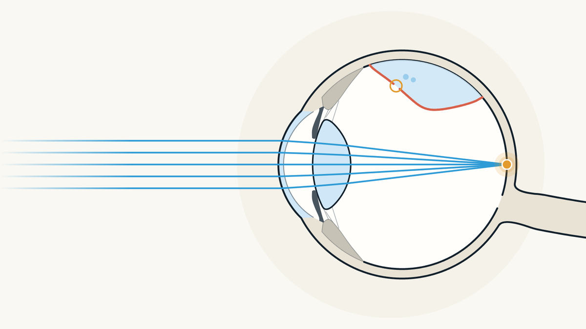
A retinal detachment is when the retina, the thin light-sensitive layer that lines the back of the eye, lifts away from the eye wall. It does not hurt, but sudden flashes, many new floaters or a shadow like a curtain are emergency signs: go to the emergency department or an eye care professional right away.
What is a retinal detachment?
It is when the retina comes away, partly or completely, from the back of the eye. The Canadian Association of Optometrists (CAO) explains that the retina is a thin layer of tissue made of cells called photoreceptors, the rods and cones, which send light signals to the brain.
Once detached, the retina stops working and vision becomes blurry, according to the American Academy of Ophthalmology (AAO). The Mayo Clinic adds that a detachment separates the retinal cells from the layer of blood vessels that brings them oxygen and nutrients. A Mayo Clinic ophthalmologist compares it to wallpaper peeling off a wall.

What are the warning signs of a retinal detachment?
Sudden flashes of light, many new floaters at once, a shadow in your side vision or a grey curtain over part of your vision. These are the four signs the AAO names; the Mayo Clinic adds blurred vision and side vision that gets worse.
A detachment is painless, the Mayo Clinic notes, and the signs often show up before it happens or gets worse. If only a small part of the retina is detached, there may be no symptoms at all, says the US National Eye Institute (NEI). To tell harmless floaters from the ones that need a prompt exam, our article on floaters and flashes goes through each case.

What does the shadow or "curtain" look like?
Like a dark area that hides part of what you see, at the side or in the middle. The NEI describes a dark shadow or "curtain" in the field of vision, and the CAO describes a drop in vision that looks like a veil coming down.
The shadow comes from the part of the retina that has detached and no longer works. According to the American Society of Retina Specialists (ASRS), it affects side vision first and can stay in place or move toward the centre; how severe the symptoms are often follows how much of the retina has detached. The NEI adds that symptoms often come on quickly.

Why is a retinal detachment an emergency?
Because the detached retina is cut off from its oxygen and the affected area can grow. The NEI explains that without prompt treatment, more of the retina can detach, which raises the risk of permanent vision loss or blindness. It advises going to your eye doctor or the emergency room right away.
The longer you wait, the less likely surgery is to succeed, the CAO stresses, and the Mayo Clinic considers surgery within days of the diagnosis ideal. Our page on urgent eye care in Montréal explains where to go for each sign: a curtain or shadow over your vision is a hospital emergency department visit.

Why does the macula matter so much?
Because it gives you sharp central vision. The AAO describes the macula as a small area in the centre of the retina that you need to see fine detail, such as faces and written text.
According to the ASRS, when the centre of the retina is still attached at the time of surgery, vision after surgery tends to stay close to what it was before. If it has detached, vision often improves, but some permanent loss may remain. In a study of 96 eyes published in 2021, final vision was poorer when the macula had been detached for 4 days or more than when it had stayed attached. An OCT scan can show whether fluid has lifted the macula, the ASRS adds.

What are the types of retinal detachment?
There are three: rhegmatogenous, tractional and exudative. The NEI explains that a rhegmatogenous detachment, the usual form, starts with a small tear: fluid from the vitreous, the gel that fills the eye, gets behind the retina and pushes it away. Its usual cause is aging, as the vitreous shrinks and can pull on the retina hard enough to tear it.
A tractional detachment happens when scar tissue on the surface of the retina pulls it away from the back of the eye. An exudative detachment comes from fluid building up behind the retina without any tear. The NEI names leaking blood vessels or swelling at the back of the eye, for example from age-related macular degeneration, inflammation, an injury or a tumour.

Who is at higher risk of a retinal detachment?
People who are nearsighted, who have had eye surgery such as cataract surgery, or who have had a serious eye injury. The AAO adds a tear or detachment in the other eye, family members who have had one, weak areas of the retina seen during an exam and some glaucoma drops that make the pupil small, such as pilocarpine.
The NEI notes that extreme nearsightedness, especially degenerative myopia, is one of the risks, and the CAO says a detachment can happen years after an eye injury or a blow to the head. According to the Mayo Clinic, it is more common between ages 40 and 70; the CAO adds that it affects men more often. Our articles on myopia and how it is corrected and on cataracts and cataract surgery explain both situations.

Can diabetes cause a retinal detachment?
Yes, mostly the tractional type. The NEI explains that diabetic retinopathy damages the blood vessels of the retina and can leave scars; as they grow, they can pull on the retina and detach it from the back of the eye. The Mayo Clinic notes that this type is usually seen when diabetes is poorly controlled.
The NEI recommends that people with diabetes get a comprehensive dilated eye exam at least once a year. It also says that managing diabetes, by staying active, eating well and taking your medicine, helps prevent or delay vision loss. The CAO also lists diabetes among the possible causes of detachment.

How is a retinal detachment diagnosed?
With an exam of the back of the eye, after the pupil is widened with drops. The NEI says this exam is usually painless and that the professional may press on the eyelids to look for a tear, which can feel a little uncomfortable.
If more information is needed, an ultrasound or an optical coherence tomography (OCT) scan shows the exact position of the retina; both tests are painless, according to the NEI. The Mayo Clinic says ultrasound is used when blood in the eye hides the retina, that both eyes are checked, and that you may be asked to come back within a few weeks if no tear is found. Our article on pupil dilation explains what the drops do.

How is a tear treated before it detaches?
With a laser or with freezing, to seal the tear before fluid gets under it. The Mayo Clinic describes laser treatment, which makes small burns around the tear, and cryopexy, a freezing probe placed on the outside of the eye right over it; both leave a scar that holds the retina against the wall.
These treatments are usually done in the office, and you go home afterwards. The Mayo Clinic advises avoiding activities that jar the eyes, such as running, for about two weeks. The CAO notes that an ophthalmologist can also treat weak areas of the retina the same way, with no detachment, to keep it from detaching.

What surgeries repair a detached retina?
There are three main ones: pneumatic retinopexy, scleral buckle and vitrectomy. In pneumatic retinopexy, the ophthalmologist injects a gas bubble into the eye that pushes the retina against the wall so it can heal, the AAO explains; the tear is sealed with freezing or a laser, the Mayo Clinic adds.
A scleral buckle is a soft band, silicone according to the Mayo Clinic, sewn onto the white of the eye to press it gently inward; you cannot see it, and it usually stays in place for good. A vitrectomy removes the vitreous that is pulling on the retina and replaces it with a bubble of air, gas or silicone oil; the oil is taken out a few months later. The choice depends on where the detachment is and how large it is, and the techniques can be combined, the Mayo Clinic notes.

What is recovery like after surgery?
Rest, an eye patch and, if a bubble was placed, a head position to hold. The AAO describes discomfort for a few days to weeks, eased with medicine, a patch to wear as long as you are told and a few weeks of lighter activity.
With a bubble, you need to keep your head in a set position, such as face down or on one side, for a period that can be 1 to 2 weeks, according to the AAO. You may see floaters, flashes and the bubble itself for a few weeks. Vision starts to improve around 4 to 6 weeks, but can keep changing for months. Your ophthalmologist tells you when you can exercise or drive again.

Can you fly with a gas bubble in your eye?
No, not until the bubble is gone. The AAO explains that pressure changes during a flight make the bubble expand, which raises the pressure in the eye and can cause severe pain and permanent vision loss. It also rules out mountains and scuba diving; your ophthalmologist confirms when the bubble has dissolved.
Nitrous oxide, a gas sometimes used in anaesthesia, must also be avoided. A report published in the BMJ in 2002 explains that it makes the bubble in the eye expand quickly, that it must not be used while the bubble is there, which can be up to three months, and that patients should know this and carry a card that says so. After a simple laser treatment for a tear, the AAO says flying is usually fine, since there is no bubble.

What are the results and risks of surgery?
Treatment is successful in the end for about 9 people out of 10, sometimes after a second procedure, according to the NEI. The AAO says about 1 patient in 20 needs more than one surgery, and that how much vision comes back depends on the damage the detachment did to the retinal cells, which can keep healing for a year or more.
Like any surgery, the repair has risks, which the AAO lists: infection, bleeding in the eye, higher pressure in the eye, cataract, a retina that does not reattach or detaches again, and permanently reduced vision. It also points out that without treatment, sight can be lost quickly and for good. The Mayo Clinic notes that some people never get all their lost vision back.

Will you need new glasses after surgery?
Possibly: your glasses prescription may change after the repair, especially with a scleral buckle, according to the Mayo Clinic. An ophthalmologist answering public questions for the AAO advises waiting until the eye has stabilized before ordering new glasses.
The Mayo Clinic recommends getting an updated prescription once the eye has healed and asking for safety lenses to protect your eyes. If some vision does not come back, it also suggests good lighting at home for reading and other everyday activities.

Can a retinal detachment be prevented, and who should get checked?
Often it cannot be prevented, because it usually comes with aging, but some risks can be lowered. The NEI and the CAO advise wearing eye protection for sports and when using power or yard tools, and the NEI recommends getting your eyes checked after any eye injury.
Regular exams can find a small tear early, before it affects your vision, according to the NEI and the CAO. If you have a risk factor, such as high myopia, a relative who had a detachment, a detachment in the other eye or past cataract surgery, the NEI suggests talking with your eye care professional about it. For people with diabetes, it recommends a dilated exam at least once a year.

Where can you have your retinas checked in Montréal?
At PrimeVision, 9520 Boulevard de l'Acadie, Suite 222, in Montréal (Ahuntsic), optometrists and a licensed optician work under one roof. The adult eye exam there includes the health of both retinas and OCT imaging. Urgent problems are seen when there is availability; otherwise, depending on how serious it is, the clinic recommends the emergency department. A detachment is repaired by an ophthalmologist: the clinic sends the referral directly and gives you a copy; wait times depend on the hospital or clinic that receives it. RAMQ covers eye exams for children under 18 and adults 65+.
If you see a curtain, a shadow, or sudden flashes and floaters, do not wait for an appointment: go to the emergency department or an eye care professional right away, as the NEI advises. For a check-up without symptoms, for example if you are very nearsighted or a relative had a detachment, go through our page to have an optometrist check the health of your retinas: book online, the times shown are the open ones.

Sources
- Detached Retina, American Academy of Ophthalmology, October 21, 2026.
- What Is Vitrectomy?, American Academy of Ophthalmology, January 22, 2026.
- Flying After Eye Surgery or With an Eye Condition, American Academy of Ophthalmology, July 6, 2026.
- Macula, American Academy of Ophthalmology, March 28, 2016.
- Will my glasses work after my retinal detachment surgery?, American Academy of Ophthalmology, April 15, 2019.
- Retinal Detachment, National Eye Institute, November 5, 2025.
- Types and Causes of Retinal Detachment, National Eye Institute, December 6, 2024.
- Retinal detachment: Symptoms and causes, Mayo Clinic, September 27, 2024.
- Retinal detachment: Diagnosis and treatment, Mayo Clinic, September 27, 2024.
- Retinal Detachment, Canadian Association of Optometrists, January 14, 2025.
- Comparison of the visual outcome between macula-on and macula-off rhegmatogenous retinal detachment based on the duration of macular detachment, Lee C.S. et al., BMJ Open Ophthalmology, 2021.
- Nitrous oxide anaesthesia in the presence of intraocular gas can cause irreversible blindness, Yang Y.F. et al., BMJ, 2002.
- Retinal Detachment, American Society of Retina Specialists, 2025 fact sheet.
Find our eye care articles useful? Add PrimeVision as a preferred source in Google.
This article is general information and does not replace an eye exam.