Visual Field Test: What It Checks and How It Feels
What the visual field is, what the test checks and why it is done, the kinds of tests, what happens step by step, why missing some lights is normal, how long it takes, how to prepare, how reliability is checked, why it is repeated, how to read the results, how often and what it means for driving.
By Michel Geha, O.O.D.
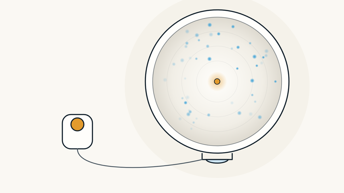
A visual field test measures the whole area each eye can see while it looks at a point straight ahead, side vision included. It finds spots where vision is weak or missing, often from glaucoma. You look at a light in the centre and press a button each time you notice a dot of light, one eye at a time.
What is the visual field?
It is the whole area one eye sees when it looks straight ahead without moving. The American Academy of Ophthalmology (AAO) defines it as how wide an area your eye can see when you focus on a central point. It includes central vision and side vision, also called peripheral vision.
Each eye is measured on its own. According to the AAO, the test shows how much vision you have in either eye and how much you may have lost over time. Since side vision can be lost without noticing, the AAO adds, this test can find problems you were not aware of.

What does a visual field test check?
It looks for spots where you cannot see, or see less, and records where they are. The AAO calls these spots scotomas. Their size and shape show how an eye disease or a brain disorder is affecting vision. Ophthalmologists also use it to measure how much a drooping eyelid blocks.
The test does not make a diagnosis on its own. For glaucoma, the clinical practice guide of the Ordre des optométristes du Québec (OOQ), the province's college of optometrists, pairs an automated visual field with eye pressure, an optic nerve exam and an OCT of the nerve fibre layer, and the results are read together.

Why would you need a visual field test?
Most often to find or follow glaucoma. The AAO also lists multiple sclerosis, thyroid eye disease, pituitary gland disorders, stroke, a tumour pressing on the parts of the brain used for vision and long-term use of certain medicines such as hydroxychloroquine (Plaquenil). Diabetes and high blood pressure can also call for it.
The Mayo Clinic explains that a large pituitary tumour can press on nearby nerves and cause loss of side vision. The OOQ guide adds other situations, such as headaches, a head injury, unexplained vision loss and age-related macular degeneration (AMD). Our article on headaches and vision explains when the eyes may be the cause.

How is the visual field linked to glaucoma?
Glaucoma damages the optic nerve, and vision loss usually starts at the sides. The US National Eye Institute (NEI) says it often affects the part of vision closest to the nose first, and moves so slowly that many people do not notice it at first. It adds that a glaucoma exam includes a visual field test to check side vision.
The AAO explains that blind spots appear as nerve fibres die, often without being noticed. Over time, sight can narrow to tunnel vision, the Canadian Association of Optometrists (CAO) notes. OCT measures the structure of the nerve and the visual field measures what it sees: the two go together. To read more, see our article on glaucoma, the one on OCT scans and our glaucoma screening and OCT page.

What kinds of visual field tests are there?
There are several, from simple to detailed. The simplest is the confrontation test: one eye is covered, you look straight ahead and the examiner holds up fingers at the sides and asks how many you see. The CAO says the examiner sits about one metre away. According to the AAO's EyeWiki, this test is poor at finding early glaucoma but can show large losses.
The automated static test, called perimetry, flashes small fixed lights of different brightness inside a bowl. A kinetic test uses a moving light instead. The AAO also describes frequency doubling perimetry, with flickering bars, and electroretinography for some retinal conditions. The Amsler grid checks only the centre of the visual field; people with AMD use it at home.

What happens during an automated test, step by step?
You sit in front of a bowl-shaped instrument with your chin on a rest and answer with a button. Here are the steps the AAO and the CAO describe:
- One eye at a time. The eye not being tested is covered with a patch.
- The right correction. Lenses matching your prescription are placed in front of the tested eye so you see as clearly as possible.
- Settled in. Your head rests on the chin rest; the CAO says the position should be comfortable.
- Eyes on the centre. You keep looking at the centre target for the whole test.
- One click per light. Small, dim lights appear around the bowl; you press the button each time you see one.
- Breaks allowed. You may blink normally and pause when needed; the CAO notes that holding the button down often does it.
The instrument records the lights you did not see. The CAO recommends a dim, quiet room, since room light and movement nearby can change the results. Then the test is repeated on the other eye.

Is it normal not to see every light?
Yes, that is by design. The AAO explains that the instrument deliberately shows lights too dim for you to see. That is how it finds your threshold at each spot, meaning the brightness you have trouble seeing half the time. Missing some lights, the AAO says, is how the test is supposed to work.
Press when you are reasonably sure you saw a light, even a faint one, without guessing. The CAO notes that some people press along with the rhythm or the sounds of the instrument; those presses with no light skew the result. So keep your eyes on the centre and answer only to what you see.

How long does a visual field test take?
A few minutes per eye, depending on the program. The AAO's EyeWiki gives about 7 minutes per eye for the SITA Standard program on Humphrey instruments, about 4 minutes for SITA Fast and about 2 minutes for SITA Faster. In a Turkish study published in 2025 of 72 people with glaucoma, average times ranged from about 3 to 7 minutes depending on the program.
Since each eye is tested separately, allow time for both eyes plus setting up. Other instruments, including Octopus perimeters, have their own programs, and a confrontation test is simple and quick. A manual kinetic test needs an experienced examiner and plotting by hand, according to EyeWiki.

How should you prepare?
Bring your glasses or a recent prescription, and come rested. The test is done with your correction in front of the eye, the AAO explains, and the CAO asks staff to record your distance prescription before starting. If drops have widened your pupils, the CAO says to wait until they are fully dilated before testing.
The test relies on your attention. A 2022 article in the Indian Journal of Ophthalmology notes that attention, motivation and fatigue can affect results, as can the wrong correction or a drooping eyelid. Speak up if the chin rest is uncomfortable or you need a break: the CAO notes that a head sitting too high or too low can create false losses at the top or bottom of the field.

How do you know a result is reliable?
The instrument checks itself during the test, with three measures described in the AAO's EyeWiki. First, fixation losses: the instrument sometimes shows a light in your blind spot; if you see it, your gaze has moved. Second, false positives: you pressed when no light appeared. Third, false negatives: you missed a brighter light at a spot where you had already seen one.
These numbers are printed with the result. According to EyeWiki, many false positives can make the map look paler than it should, while false negatives often point to a lapse in attention. The CAO advises staff to pause and go over the instructions again when these errors add up.

Why is the test sometimes repeated?
Because people get better with practice, and one test is not enough to follow a disease. In a 1996 study by Heijl and Bengtsson, 25 people newly diagnosed with glaucoma took the test five times about a week apart. Results improved in most of them, mainly between the first and second test.
The authors conclude that a baseline for follow-up should include more than one test. For glaucoma follow-up, a 2025 Australian study refers to a recommended 6 tests in the first 2 years. A weaker first result can therefore improve on the next test, and change is judged over the series.

What do the results look like?
A map of each eye, with numbers. The AAO's EyeWiki explains that the report gives the sensitivity measured at each point, a grey-scale map where less sensitive areas look darker, and a comparison with people of the same age who see normally. It also shows the date, the correction used and the reliability measures.
A dark spot near the centre, on the temple side, is normal. The AAO explains that everyone has a blind spot where the optic nerve connects to the retina, because there are no light-sensitive cells there. EyeWiki places it about 10 to 15 degrees from the centre. A cataract or a small pupil can also darken the whole map, EyeWiki adds.


How often should the test be done?
It depends on your situation, and the professional following you decides. The OOQ guide calls for a visual field screening at an adult's first exam and at every exam from age 40, and earlier when there are risk factors. It adds that your history and what the exam shows set the type of test and how often it is repeated.
With glaucoma, the OOQ calls for a visual field at screening and then periodically during follow-up. For hydroxychloroquine, the AAO mentions a yearly visual field check, and the OOQ a yearly exam after 5 years of use, or sooner with risk factors. People with AMD use the Amsler grid once a day, every day, according to the AAO.

Does the visual field matter for driving?
Yes. The AAO warns that a limited visual field can put your ability to drive at risk, and advises talking to your eye doctor if you are concerned. The CAO notes that the tunnel vision of advanced glaucoma can interfere with activities such as driving.
The OOQ guide lists a driver's licence form among the situations where a specific visual field study is recommended. The exact licence requirements are not covered here: if you have a form to fill out, bring it and ask about it during the exam.

Where can you have your visual field checked in Montréal?
At PrimeVision, 9520 Boulevard de l'Acadie, Suite 222, in Montréal (Ahuntsic), optometrists and a licensed optician work under one roof. The clinic has a Haag-Streit Octopus 900 for visual field testing and a Canon RS-330 OCT for retinal imaging. The optometrist decides whether you need a visual field test. If an ophthalmologist's opinion is needed, the clinic sends the referral directly and gives you a copy; wait times depend on the hospital or clinic that receives it and can be long. RAMQ covers eye exams for children under 18 and adults 65+.
To book, go through our page to see how a full eye exam goes at the clinic. Book online: the times shown are the open ones.

Sources
- Visual Field Test and Blind Spots (Scotomas), American Academy of Ophthalmology, March 5, 2025.
- Understanding Glaucoma, American Academy of Ophthalmology, March 4, 2026.
- Eye Exam and Vision Testing Basics, American Academy of Ophthalmology, February 14, 2024.
- All About the Amsler Grid and Daily Vision Checks for AMD, American Academy of Ophthalmology, December 19, 2025.
- Experiment: Blind Spot, American Academy of Ophthalmology, accessed November 2, 2026.
- Standard Automated Perimetry, EyeWiki, American Academy of Ophthalmology, last edited November 2, 2026.
- Glaucoma, National Eye Institute, August 19, 2026.
- Pituitary tumors and adenomas, Mayo Clinic, accessed November 2, 2026.
- Glaucoma, Canadian Association of Optometrists, accessed November 2, 2026.
- Automated Perimetry, CCOA Program, Module 3, Canadian Association of Optometrists, July 22, 2019.
- Confrontation Field Screening, CCOA Program, Module 3, Canadian Association of Optometrists, July 22, 2019.
- Guide d'exercice clinique, Ordre des optométristes du Québec, February 20, 2023 (in French).
- Heijl A, Bengtsson B. The effect of perimetric experience in patients with glaucoma, Archives of Ophthalmology, January 1996.
- Köksaldı S et al. Comparison of Humphrey 24-2 SITA Standard, SITA Fast, and SITA Faster Test Strategies in Patients with Glaucoma, Turkish Journal of Ophthalmology, April 24, 2025.
- Wang H et al. The Frontloading Approach to Meet Guideline-Recommended Visual Field Testing for Glaucoma, Ophthalmology Glaucoma, 2025.
- Mansoori T. Red flags and artifacts in perimetry, Indian Journal of Ophthalmology, December 2022.
Find our eye care articles useful? Add PrimeVision as a preferred source in Google.
This article is general information and does not replace an eye exam.