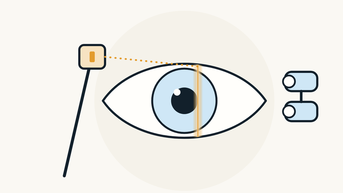
Macular Degeneration (AMD): Early Signs and the Amsler Grid
Age-related macular degeneration explained: what it does to central vision, the early signs by stage, dry and wet AMD, risk factors, how to use the Amsler grid at home, when to see an eye doctor right away, how it is found with OCT, the treatments, and the habits linked to a lower risk.
By Michel Geha, O.O.D.
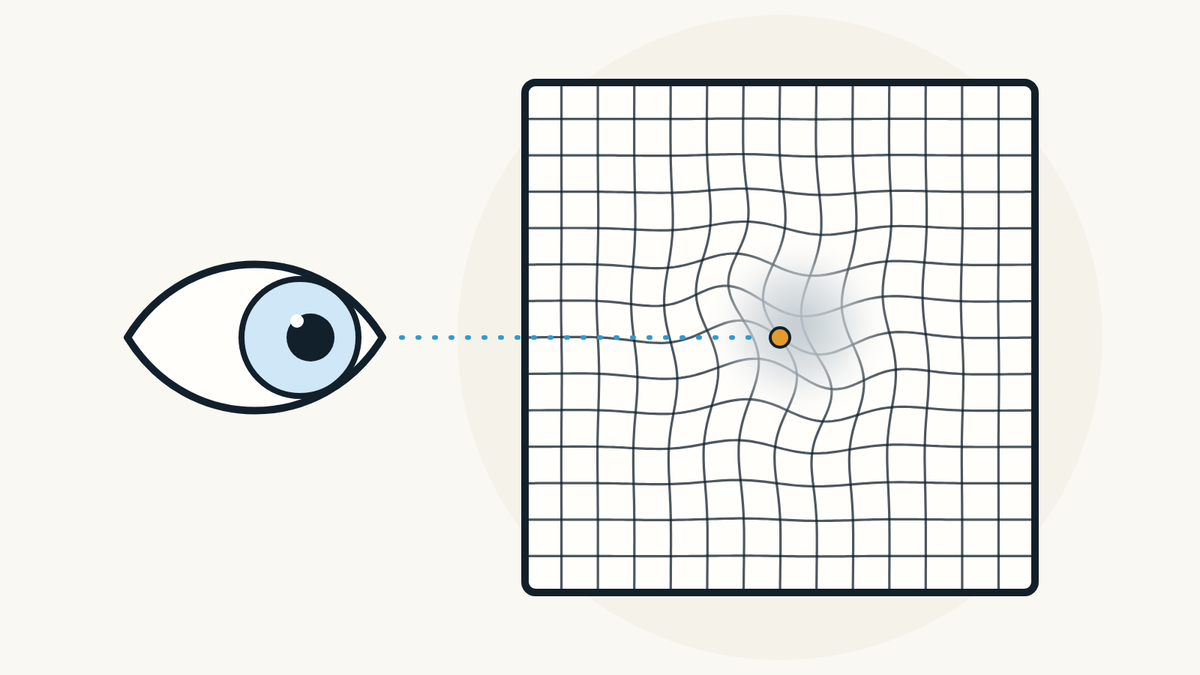
Age-related macular degeneration (AMD) is a disease of the retina that blurs central vision, the vision you use to read and recognize faces. Early on it often causes no symptoms. The first signs people notice are straight lines that look wavy and a blurry spot in the centre of their vision.
What is macular degeneration?
It is an eye disease that damages the macula with age. The US National Eye Institute (NEI) explains that the macula is part of the retina, the light-sensitive tissue at the back of the eye, and that it gives sharp, straight-ahead vision. AMD is a leading cause of vision loss in older adults.
The American Academy of Ophthalmology (AAO) gives a simple picture: looking at a clock with hands, a person with AMD may see the numbers around the edge but not the hands in the middle. AMD does not affect side vision, and according to the NEI it does not cause complete blindness.

What are the early signs of AMD?
At the very start, there are often none. The NEI says early dry AMD causes no symptoms. In the intermediate stage, some people still have none; others notice mild blur in their central vision or trouble seeing in low light.
In late AMD, wet or dry, many people see straight lines turn wavy or crooked. A blurry area may appear near the centre, grow over time or leave blank spots, and colours seem less bright. The NEI calls wavy lines a warning sign: see an eye doctor right away. If your vision is blurry for another reason, our article on blurry vision covers the common causes.

Dry or wet AMD: what is the difference?
The dry form is slow and common; the wet form is rarer and faster. According to the AAO, about 8 in 10 people with AMD have the dry form: the macula thins with age and small deposits called drusen build up under the retina. The NEI describes three stages of dry AMD, early, intermediate and late, which often progress over several years.
The wet form happens when new, abnormal blood vessels grow under the retina. They can leak blood or fluid and scar the macula, which causes faster vision loss. The NEI adds that any stage of dry AMD can turn into wet AMD.

Who is more at risk?
Risk rises with age. The AAO speaks of people over 50; the NEI of those 55 and older. Both name a family history of AMD and smoking, and note that white people are at higher risk.
The AAO adds high blood pressure, being overweight, a diet high in saturated fat (meat, butter, cheese), heart disease and high cholesterol. If you are in one of these groups, the NEI advises regular eye exams and asking your doctor how often to have them. Our article on how often to have an eye exam by age gives the Canadian guidance.

How do you use an Amsler grid?
You look at the dot in the centre of a grid, one eye at a time, and notice whether any lines change. The AAO describes the Amsler grid as a square grid with a dot in the middle, and advises people with AMD to use it once a day, every day, because vision often fades so slowly that people do not notice.
- Get ready. Put on your reading glasses if you use them and sit in good light.
- Hold the grid 12 to 15 inches (30 to 38 cm) from your face.
- Cover one eye and look at the centre dot with the other.
- Keeping your eye on the dot, notice whether all the lines look straight, or whether any area looks blurry, wavy, dark or blank.
- Repeat with the other eye.
The AAO suggests keeping the grid somewhere you see every day, such as the fridge door or the bathroom mirror. It does not replace an exam: it helps you notice a change between visits.

When should you see an eye doctor right away?
As soon as any part of the grid, or of what you look at, becomes wavy, blurry, dark or blank. The AAO advises calling your eye doctor right away in that case, and the NEI says the same about straight lines that look wavy. The AAO explains that treatment started early can help slow or stop further vision loss.
The NEI also notes that late AMD in one eye raises the risk of late AMD in the other. A good eye can hide a change in the other one: that is why each eye is tested on its own.

How does an optometrist check for AMD?
With a comprehensive eye exam that includes a look at the back of the eye. According to the NEI, drops are used to widen the pupil, then the retina is checked; the doctor may also recommend optical coherence tomography (OCT), which takes pictures of the inside of the eye. The AAO adds the Amsler grid during the exam.
OCT shows the macula in cross-section, layer by layer, which helps reveal drusen or abnormal vessels. Our article on the OCT scan explains what the image shows, and the one on pupil dilation covers what to plan when drops are used. If the wet form is suspected, the AAO says a fluorescein angiogram may be done to see the retina's blood vessels.

Can macular degeneration be treated?
Some forms can, and the ophthalmologist decides the treatment. The NEI explains that there is no treatment for early AMD: it is followed with regular exams. In the intermediate stage, specific supplements, the AREDS2 formula, may stop it from turning into late AMD.
For wet AMD, the NEI lists anti-VEGF medicines injected into the eye and photodynamic therapy, which combines an injection and a laser. The AAO adds that injections can slow geographic atrophy, a late form of dry AMD. According to the NEI's AREDS studies, these supplements do not prevent AMD from starting, and current and former smokers should avoid the formula with beta-carotene. Our article on eye vitamins sums up what those studies showed. Talk to your doctor before starting a supplement.

Can you lower your risk of AMD?
Yes, some habits are linked to a lower risk. The NEI says research shows you may lower your risk of AMD, or slow vision loss from it, by quitting smoking, getting regular physical activity, keeping blood pressure and cholesterol healthy, and eating leafy green vegetables and fish.
The AAO also advises protecting your eyes from UV light. Our article on sunglasses and UV protection explains what UV400 means.

How do you live with central vision loss?
With low vision aids and rehabilitation. The NEI points out that not everyone with AMD reaches the late stage or has it in both eyes. For those who lose central vision, low vision devices and vision rehabilitation services help people stay independent.
The AAO lists different kinds of magnifiers and electronic devices, and explains that a vision rehabilitation specialist can teach you to use your side vision for everyday tasks. Ask your eye doctor to point you to these services.

Where can you have your retina checked in Montréal?
At PrimeVision, 9520 Boulevard de l'Acadie, Suite 222, in Montréal (Ahuntsic), optometrists and a licensed optician work under one roof. The eye exam for adults 18 to 64 there includes vision and prescription tests, the health of both retinas and OCT imaging, done with a Canon RS-330; the visual field is measured with a Haag-Streit Octopus 900. If an exam calls for an ophthalmologist's opinion, the clinic sends the referral itself and gives you a copy; the wait depends on the hospital or clinic that receives it. RAMQ covers eye exams for children under 18 and adults 65+, and we see children from age 4.
Our page on glaucoma screening and OCT imaging in Montréal describes these tests, and the eye exam in Montréal page explains what the exam includes. To book, book ahead online: the times shown are the open ones. Other eye conditions are gathered in eye conditions from A to Z.

Sources
- Age-Related Macular Degeneration (AMD), National Eye Institute, updated June 22, 2021, accessed November 6, 2026.
- Understanding Macular Degeneration, American Academy of Ophthalmology, October 1, 2024, accessed November 6, 2026.
- Have AMD? Save Your Sight with an Amsler Grid, American Academy of Ophthalmology, May 3, 2023, accessed November 6, 2026.
- AREDS/AREDS2 Clinical Trials: major findings, National Eye Institute, accessed November 6, 2026.
Find our eye care articles useful? Add PrimeVision as a preferred source in Google.
This article is general information and does not replace an eye exam.