The Slit Lamp Exam: What the Optometrist Looks At
The slit lamp exam explained: what the instrument is, how the exam goes, what the optometrist checks from the eyelids to the lens, why a yellow fluorescein drop is used, how the retina and optic nerve can be seen, whether it hurts, and what to plan if your pupils are dilated.
By Michel Geha, O.O.D.
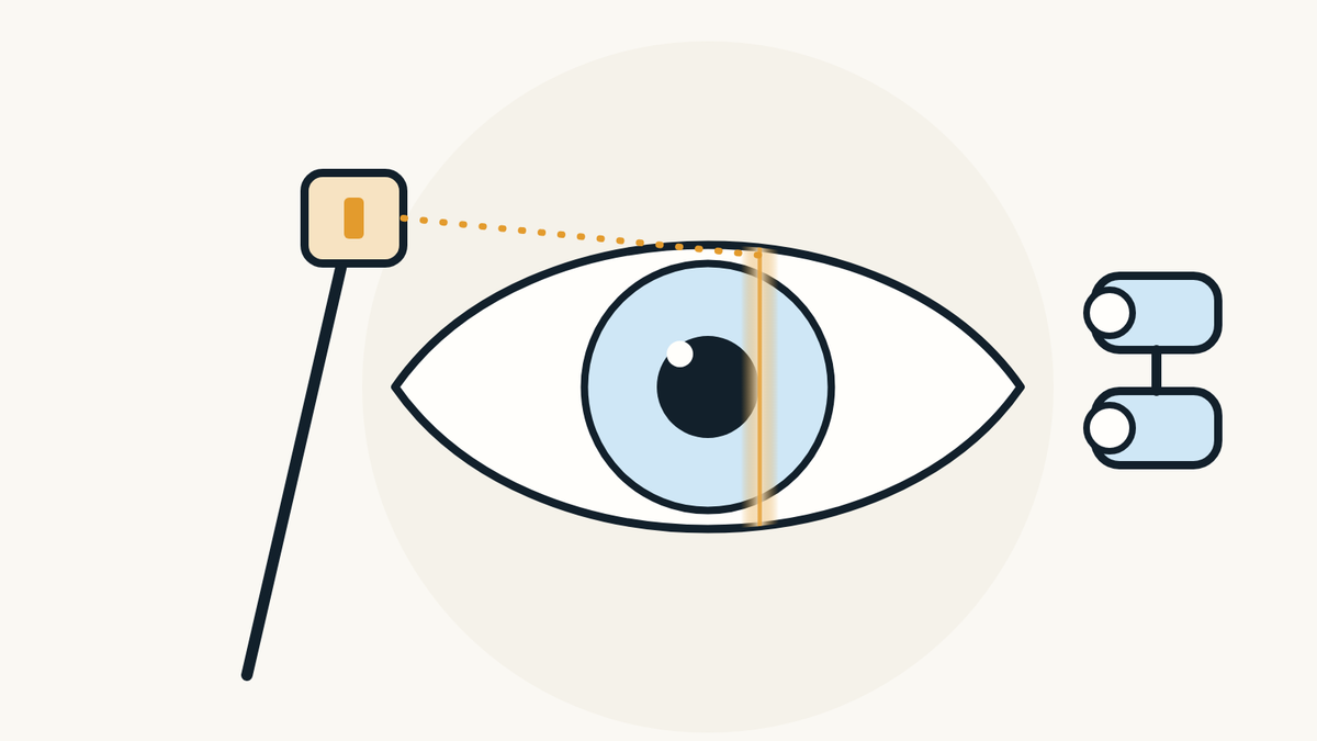
The slit lamp exam is the part of the eye exam where you rest your chin on a support and the optometrist looks at your eyes through a microscope, using a thin beam of light. It checks the health of the eye, from the eyelids to the lens, and sometimes as far back as the retina.
What is a slit lamp exam?
It is an exam of your eyes under a microscope, lit by a bright light. According to the American Academy of Ophthalmology (AAO), the slit lamp gives a closer look at the structures at the front of the eye and inside the eye, and it is a key tool for judging eye health and detecting eye disease.
The name comes from its beam. EyeWiki, the AAO's encyclopedia, describes it as a microscope that projects a beam whose height, width and angle can be adjusted. This slit of light shows the eye in depth and in cross-section, a bit like a thin slice through the clear tissues.

How does the exam go?
You sit in front of the instrument with your chin on the chin rest and your forehead against a band. The AAO explains that this position keeps your head steady, then the examiner sits facing you, looks through the microscope and aims a narrow beam of light at your eye.
According to EyeWiki, the right eye is usually examined first, then the left. You may be asked to look up, down or to the side, so that each part of the eye passes through the beam.

What is checked at the front of the eye?
The eyelids, lashes, white of the eye, cornea, iris and lens. EyeWiki describes an exam done in a set order: the structures around the eye, the lids and lashes, the conjunctiva and sclera (the white of the eye), the cornea, the anterior chamber (the space between the cornea and the iris), the iris and pupil, then the lens.
The AAO notes that this part of the exam checks for cataracts and for scars or scratches on the cornea. With a thin, angled beam, the cornea appears in cross-section, which helps place a problem within its thickness.

Why are yellow drops put in the eye?
To make damaged areas of the cornea stand out. The AAO explains that a drop with a yellow dye, fluorescein, helps find an injury such as a scratch on the cornea (corneal abrasion), or an infection such as herpes keratitis.
The dye is used with the slit lamp's blue filter: according to EyeWiki, under this blue light an abrasion or an ulcer shows up clearly.

What problems can the exam show?
Many problems at the front of the eye can be seen with a slit lamp. According to the AAO, it can show signs of dry eye, pink eye from an allergy or infection, inflammation of the white of the eye, abnormal deposits in the cornea, and cataracts.
The eyelid margins matter too. EyeWiki lists signs of blepharitis on the lashes, as well as a stye or chalazion. A sign seen at the exam is not a diagnosis on its own: your optometrist reads it together with your symptoms and the rest of the exam.

Can a slit lamp see the back of the eye?
Yes, with an extra lens. EyeWiki explains that with handheld lenses, the examiner can also view the back of the eye. The AAO adds that with fully dilated pupils, the retina and optic nerve can be seen.
According to the AAO, this helps find a torn or detached retina, and it is part of diagnosing glaucoma and age-related macular degeneration. The drops that widen the pupil are explained in our article on pupil dilation.

Does the exam hurt?
No. The AAO says that although the light is very bright, it will not damage your eye and should not cause pain. According to EyeWiki, the risks of the exam are minimal.
If you are sensitive to light or your eye is sore, say so: EyeWiki notes that the examiner can lower the brightness or use a filter, and keep the exam short.

Do you need to prepare, and can you drive after?
No special preparation is needed, according to the AAO. Still, bring your glasses, and your contact lenses or their boxes if you wear them. If your pupils are dilated, your vision will be blurry and your eyes very sensitive to light for several hours.
In that case, the AAO advises not driving after the exam, bringing sunglasses and having someone drive you home. The drops usually take 20 to 30 minutes to fully open the pupil, according to the AAO.

Is the slit lamp part of a complete eye exam?
Yes. EyeWiki calls it a mainstay of the comprehensive eye exam, letting optometrists and ophthalmologists assess every part of the eye. The AAO lists it among the steps of the exam, along with vision, pupils, side vision, eye pressure, the retina and the optic nerve.
It adds to the other tests without replacing them. Pressure is explained in our article on the eye pressure test, the prescription in our article on the refraction test, and retinal imaging in our article on the OCT scan.

Where can you have a complete eye exam in Montréal?
At PrimeVision, 9520 Boulevard de l'Acadie, Suite 222, in Montréal (Ahuntsic), optometrists and a licensed optician work under one roof. The adult eye exam there includes vision and prescription tests, the health of both retinas and OCT imaging (Canon RS-330); eye pressure and autorefraction are measured with a Nidek Tonoref III. RAMQ covers eye exams for children under 18 and adults 65+, and we see children from age 4.
The eye health check is part of the eye exam in Montréal. To book, book ahead online: the times shown are the open ones.

Sources
- What is a Slit Lamp?, American Academy of Ophthalmology, accessed November 5, 2026.
- Eye Exam and Vision Testing Basics, American Academy of Ophthalmology, accessed November 5, 2026.
- What Are Dilating Eye Drops?, American Academy of Ophthalmology, accessed November 5, 2026.
- Slit Lamp Examination, EyeWiki, American Academy of Ophthalmology, reviewed June 10, 2026, accessed November 5, 2026.
Find our eye care articles useful? Add PrimeVision as a preferred source in Google.
This article is general information and does not replace an eye exam.